

A published literature review has highlighted the need for increased awareness of ocular risks in aesthetic practice; collaboration between aesthetic practitioners and ophthalmologists is helping to improve patient safety.
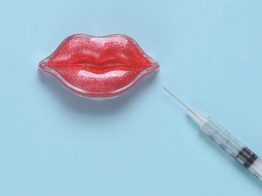
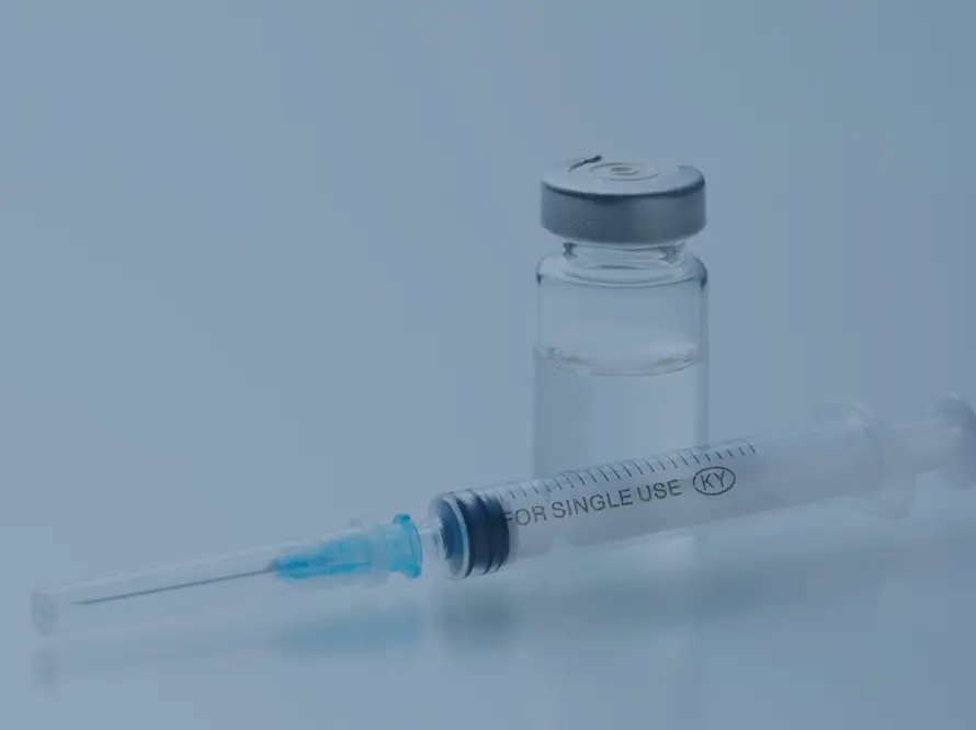
Facial aesthetic treatments, predominantly delivered as injectables, such as dermal fillers, botulinum toxins, biostimulators, skin boosters, PRP, etc., are the mainstay of aesthetic practice. Generally considered safe in appropriately trained and medically competent hands, where adherence to safer injection techniques, correct product placement, suitable patient selection, and informed consent are undertaken, such procedures do still carry the risk of causing serious ophthalmological complications, including permanent blindness from dermal filler embolisation or optic nerve damage at their most catastrophic.
The incidence of the most severe eye complications related to vision loss is low. Still, other ocular risks, including milder, transient adverse effects such as droopy eyelids from ptosis or dry eye after botulinum toxin, can be very distressing for both the patient and the practitioner, making awareness and prevention of ocular complications a core component of education for any practising aesthetic clinician.
Let’s take a deeper dive into a recently published narrative review that summarised the incidence, clinical features, risk factors, diagnostic approaches, and management strategies for several ocular complications associated with medical aesthetic treatments.[i]
Iatrogenic ocular complications in aesthetic medicine are not new
We have been discussing the potential for filler-induced blindness via ophthalmic artery/central retinal artery occlusion through the inadvertent intravascular injection of dermal filler products into their anastomoses for some considerable time. Most notably, it was first brought to widespread awareness within the aesthetic sector thanks to a clinical paper review by Lazzeri et al. in 2012, where the injection of various agents, including hyaluronic acid, silicone oil, and autologous fat, was the cause of irreversible vision loss.[ii] Since then, numerous studies have explored the risk of blindness from cosmetic injections and evaluated documented adverse events.
Reported and published patient cases of vision loss through cosmetic filler emboli are thankfully low in number, just under 200 to date, based on literature reviews. It is estimated at a risk of 1 in 100,000 dermal filler injections. However, 83% were attributed to hyaluronic acid-based products, and the nose was the most prevalent original injection site, in 40% of cases. Significantly, the vision loss in 70% of patients within the reviewed reports remained unchanged, without improvement, despite varying medical interventions.[iii] Although smaller in number, several cases have also been reported of irreversible blindness due to iatrogenic occlusion of the ophthalmic artery following Platelet-Rich-Plasma (PRP) treatment, including four patients following PRP injections in the glabella and nasolabial folds.[iv]
Ocular complications can be categorised by their nature (vascular, motor, or surface). And whilst botulinum toxin usually causes self-limiting, temporary problems, dermal fillers can have more catastrophic outcomes with few guaranteed ‘antidotes’ for resolution, making prevention better than cure.
Other reported adverse ocular effects associated with aesthetic treatments and their estimated frequency include:
- Temporary palpebral ptosis or a transient droopy eyelid from botulinum toxin diffusion to the levator muscle, considered a common complication, resolving after a couple of months.
- Orbital cellulitis and periocular infection are relatively frequent, with an estimated 6% risk of occurrence; they are managed with antibiotics and monitoring.
- Temporary dry eye due to incomplete eyelid closure and blinking following botulinum toxin injection to the orbicularis oculi muscle, estimated at a risk of 3% frequency, resolves with muscle recovery; symptoms can be treated with artificial tears and overnight lid closure.
- Oculomotor nerve palsies or muscle dysfunction (e.g., diplopia or double vision from botulinum toxin diffusion to the lateral rectus muscle or orbital ischemia from dermal fillers); usually resolves spontaneously, has up to a 2.2% risk of occurrence.
- Periocular skin necrosis that can lead to exposure keratopathy or damage to the cornea from incomplete eyelid closure and eye dryness due to scarring and tissue contraction affecting the eye; a rare yet reported occurrence.
- Ischemic optic neuropathy or optic nerve damage usually happens alongside arterial occlusion of the retina from dermal filler embolisation; it has only been reported in some isolated cases.
- Other periocular tissue reactions, including oedema, hematoma, granuloma etc.
Clinicians have also questioned whether a low concentration of naturally present or endogenous hyaluronidase in the tear trough area, under the eye, explains the incidences of prolonged filler presence (due to slow degradation from the lack of natural hyaluronidase), persistent oedema, or unusual and often delayed reactions after periocular dermal filler treatment. Current, published clinical literature does not provide direct answers or outcomes to support this hypothesis, but studies such as those published by Mobin Master et al. do demonstrate that hyaluronic acid-based products persist longer than claimed, particularly in less mobile areas of the face.[v],[vi],[vii]
Mitigating ocular complications when delivering cosmetic injectables
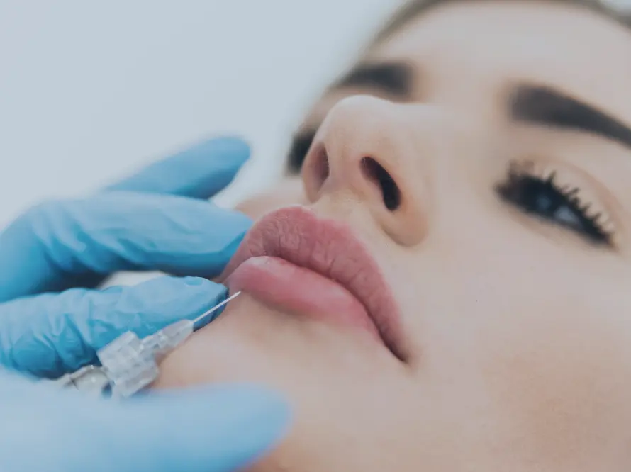
A greater understanding of facial anatomy, and most importantly, facial vasculature, is the foundation for preventing ocular complications. Alongside consideration of safer injection techniques using cannulas rather than sharp needles, for example, with slow injections and micro bolus deposits, plus an awareness, caution, and potential avoidance of so-called “danger zones”, most notably the nasal dorsum (bridge of the nose) and glabella.
Many facial regions have direct circulatory connections through vascular anastomoses to the ophthalmic artery (and the central retinal artery), meaning accidental intravascular deposition has a seamless route to the eye. These routes emanate from the glabellar area supplied by the supratrochlear artery, the dorsal nasal artery, the forehead and supraorbital artery, and the territory of the angular artery near the nose or medial canthus. Injections in these areas, therefore, carry a greater risk. Mitigation and prevention of complications by understanding these “danger zones”, an emergency kit, protocol, and plan for specialist referral are paramount.
Patients should routinely be made aware of the potential for any number of ocular complications with injectable aesthetic treatments, including the risk of blindness, no matter how rare, as part of the process of seeking informed consent for treatment. A lack of disclosure carries ethical, medicolegal, and insurance implications. They should be instructed to report any impaired visual symptoms immediately – during or after injections – to avoid delayed recognition of a problem, which can contribute to a worse outcome.
As aesthetic medical practitioners and not ophthalmologists, one obstacle is recognising and identifying the signs of an ocular complication, alongside appropriate (emergency) management, which should include pre-determined referral pathways to specialist eyecare services and physicians, should the worst happen.
The onset of vision changes (usually to one eye) in cases of arterial occlusion is immediate, during the injection or a few seconds afterwards, combined with severe eye pain. Associated symptoms include difficulty moving their eye, ptosis of the eyelid, skin discolouration and blanching nearby, headache, dizziness, and nausea. Sadly, effective treatment is usually limited for vision prognosis, but prompt treatment (which can include retrobulbar hyaluronidase for hyaluronic acid-based products and hyperbaric oxygen therapy), plus assistance and guidance from ocular specialists, should be sought.
Consider strengthening your relationships and networks to include an ophthalmological specialist in your local area for emergency referral, training and shared learning as part of a multidisciplinary approach to your aesthetic practice and patient safety. Think about joining aesthetic complications groups available to medical practitioners. Many offer educational events and online resources, emergency support services, and peer-based communities for continued learning surrounding complications from aesthetic treatments.
References
[i] De-Pablo-Gómez-de-Liaño L, Ly-Yang F, Burgos-Blasco B, Fernández-Vigo JI. Ophthalmological Complications of Aesthetic Medicine Procedures: A Narrative Review. J Clin Med. 2025 Jul 31;14(15):5399. doi: 10.3390/jcm14155399. PMID: 40807018; PMCID: PMC12347381. https://pmc.ncbi.nlm.nih.gov/articles/PMC12347381/
[ii] Lazzeri D, Agostini T, Figus M, Nardi M, Pantaloni M, Lazzeri S. Blindness following cosmetic injections of the face. Plast Reconstr Surg. 2012 Apr;129(4):995-1012. doi: 10.1097/PRS.0b013e3182442363. PMID: 22456369. https://pubmed.ncbi.nlm.nih.gov/22456369/
[iii] Foster J, Aakalu VK, Freitag SK, McCulley TJ, Tao JP, Vagefi MR, Yen MT, Kim SJ, Wladis EJ. Vision-Threatening Complications of Soft Tissue Fillers: A Report by the American Academy of Ophthalmology. Ophthalmology. 2025 Aug;132(8):935-944. doi: 10.1016/j.ophtha.2025.01.020. Epub 2025 Mar 31. PMID: 40167411. https://www.aaojournal.org/article/S0161-6420(25)00074-0/fulltext
[iv] Karam EZ, Gan A, Muci Mendoza R, Martinez E, Perez E. Visual Loss after Platelet-rich Plasma Injection into the Face. Neuroophthalmology. 2020 Mar 26;44(6):371-378. doi: 10.1080/01658107.2020.1740936. PMID: 33335344; PMCID: PMC7722697. https://pmc.ncbi.nlm.nih.gov/articles/PMC7722697/
[v] Master M. Hyaluronic Acid Filler Longevity and Localization: Magnetic Resonance Imaging Evidence. Plast Reconstr Surg. 2021 Jan 1;147(1):50e-53e. doi: 10.1097/PRS.0000000000007429. PMID: 33002985. https://pubmed.ncbi.nlm.nih.gov/33002985/
[vi] Master M, Azizeddin A, Master V. Hyaluronic Acid Filler Longevity in the Mid-face: A Review of 33 Magnetic Resonance Imaging Studies. Plast Reconstr Surg Glob Open. 2024 Jul 15;12(7):e5934. doi: 10.1097/GOX.0000000000005934. PMID: 39015357; PMCID: PMC11250456. https://pmc.ncbi.nlm.nih.gov/articles/PMC11250456/
[vii] Master M, Roberts S. Long-term MRI Follow-up of Hyaluronic Acid Dermal Filler. Plast Reconstr Surg Glob Open. 2022 Apr 13;10(4):e4252. doi: 10.1097/GOX.0000000000004252. PMID: 35433153; PMCID: PMC9007185. https://pmc.ncbi.nlm.nih.gov/articles/PMC9007185/
Categories
Related news articles